
B-Scan Evaluation of Medial and Lateral Rectus Muscle Thickness in Horizontal Strabismus
Doi: 10.36351/pjo.v42i2.2224
DOI:
https://doi.org/10.36351/pjo.v42i2.2224Abstract
Purpose: To evaluate the Medial and Lateral Rectus Muscle Thickness in Horizontal Strabismus.
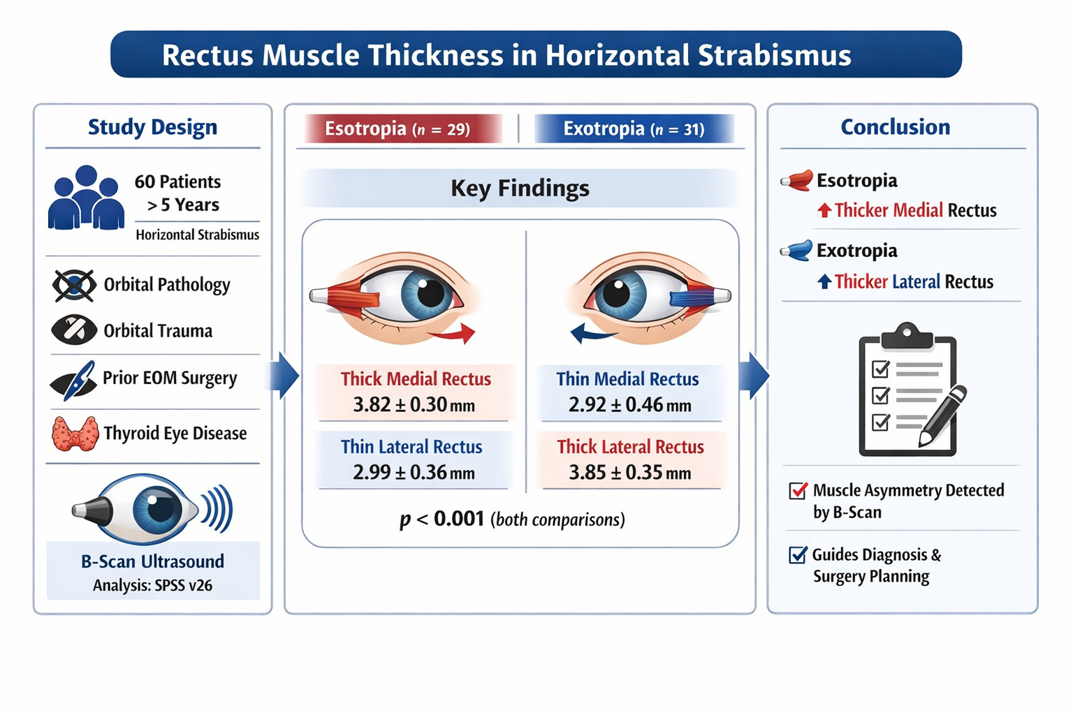
Study Design: Descriptive observational study.
Place and Duration of Study: University of Lahore hospital, Lahore from July 2025 to October 2025.
Methods: Sixty patients, above 5 years of age with horizontal strabismus were included. Patients with orbital pathology, orbital trauma, previous surgery of extraocular muscles and Thyroid eye disease were excluded. After complete history and examination, B-scan was done to evaluate medial and lateral rectus muscles. IBM SPSS version 26 was used for data analysis.
Results: There were 22 males and 38 females. The mean age was 12.5±1.8 years. Twenty-nine patients had esotropia and 31 had exotropia. Medial rectus muscle was significantly thicker in eyes with esotropia compared to those with exotropia (Mean of 3.82 ± 0.30mm vs. 2.92 ± 0.46 mm; p < 0.001). In contrast, the lateral rectus muscle was significantly thinner in esotropic eyes than in exotropic eyes (2.99 ± 0.36 mm vs. 3.85 ± 0.35 mm; p < 0.001). These findings demonstrate a clear and statistically robust pattern of rectus muscle asymmetry between the two forms of horizontal strabismus.
Conclusion: B-scan ultrasonography showed that esotropia was associated with a significant increase in medial rectus thickness, whereas exotropia was associated with increase in lateral rectus thickness. Knowledge of muscle thickness can assist surgeons in tailoring the amount of recession or resection. Thicker or hypertrophied muscles may respond differently to standard surgical resection or recessions.
Downloads
Published
How to Cite
Issue
Section
License
Copyright (c) 2026 Tahir Shaukat, Rashida Riaz, Muhammad Hannan Jamil, Dr. Khalid Rafique, Dr. Humera Zafar Ali

This work is licensed under a Creative Commons Attribution-NonCommercial 4.0 International License.


